Sclerotherapy sits at an interesting intersection of cosmetic improvement and medical care. It can make legs look clearer and feel lighter, yet it is also a practical therapy for venous insufficiency in the right patients. After treating thousands of veins under magnification and ultrasound, I can tell you that sclerotherapy is not one thing. It changes character depending on the vein you are treating. A tiny spider vein on the ankle behaves nothing like a ropey varicose vein on the calf. Understanding those differences helps set expectations for sclerotherapy results, recovery, and how many sessions it might take.
This guide looks at how sclerotherapy works, why spider veins respond so well, when varicose veins need foam or ultrasound guidance, and what influences the durability of results. I will weave in practical detail from the clinic floor so you know what to ask and how to prepare.
How sclerotherapy works in plain terms
Sclerotherapy is an injection therapy. A clinician introduces a sclerosant solution into a vein to irritate its inner lining. That irritation prompts the vein walls to stick together, collapse, and eventually scar down. Blood reroutes to healthier veins. The body then slowly resorbs the treated vein over weeks to months.
There are two broad forms. Liquid sclerotherapy uses a low to moderate concentration of solution, typically for fine veins close to the skin. Foam sclerotherapy mixes the sclerosant with air or physiologic gas to create microbubbles, which push out blood and increase contact with the vein wall. Foam works better for larger, slower-flowing veins like reticular and varicose veins, and it shows up clearly on ultrasound.
Common agents include polidocanol and sodium tetradecyl sulfate. Concentration, volume per injection, and spacing between injections all vary by vein size, location, and the patient’s history. The sclerotherapy process is brief, usually an office-based treatment lasting 15 to 45 minutes depending on the number of veins addressed. Most patients resume normal activities the same day, with walking encouraged.
Spider veins versus varicose veins: what you are really treating
Spider veins, the technical term is telangiectasias, are small red, purple, or blue lines that sit right under the skin. They average 0.1 to 1 millimeter in diameter. They can map out like a fan around the ankle or sprawl across the outer thigh. Spider veins may itch or sting, but many people seek sclerotherapy for cosmetic reasons.
Varicose veins are bigger, more than 3 millimeters, and often bulge or twist. They connect to a deeper network and frequently derive from valve incompetence in a saphenous trunk or a perforating vein. These veins can ache, throb, cramp at night, and swell with prolonged standing. Left unchecked, severe venous insufficiency can lead to skin changes and ulceration, especially around the inner ankle.
Reticular veins sit in the middle. Think of them as feeder veins, 1 to 3 millimeters deep blue or green lines, often supplying clusters of spider veins from underneath. Reticular veins matter because treating them first makes spider vein sclerotherapy more effective and reduces recurrence.
Why spider veins usually respond better and faster
If a patient asks, will sclerotherapy get rid of my spider veins, the honest answer is usually yes, though it may take more than one session. Spider veins are small targets, and liquid micro sclerotherapy gets the job done. When the feeder vein is addressed and compression is used diligently after treatment, clearance rates for spider veins often exceed 70 to 80 percent after one to three sessions. The skin looks more uniform, and photos taken at six to twelve weeks show meaningful change.
There are caveats. Ankle and foot telangiectasias are slower to fade and prone to matting, those tiny red networks that can form after treatment. Darker skin types can develop hyperpigmentation for a time, especially if the vein was larger or there was bruising. Fragile, sun-damaged skin may need lower concentrations and gentle technique. Hormonal changes can fuel new spider veins even after perfect treatment, which is why maintenance touch-ups every 1 to 3 years are common for those who care about appearance.
A clinical detail that helps: addressing reticular feeders first. If you see a blush of spiders radiating from a specific region, ultrasound or transillumination may reveal a 2 millimeter reticular vein beneath. Treating that vessel first reduces pressure and makes the more superficial injections more durable. Patients notice fewer “reappearing” veins when the feeder is handled.
Varicose veins need a different playbook
When patients arrive with ropey, bulging veins and throbbing after a long shift on their feet, we start with an ultrasound. Sclerotherapy can be effective for varicose veins, but only when used in the right context. If the great saphenous vein or small saphenous vein is severely incompetent, treating only surface varicosities is like bailing water without plugging the leak. In those cases, the best path is to close the faulty trunk first. That may be done with endovenous laser ablation, radiofrequency ablation, cyanoacrylate closure, or occasionally foam sclerotherapy under ultrasound.
Once the refluxing trunk is addressed, foam sclerotherapy shines for branch varicosities and residual clusters. Under ultrasound guidance, we can map the pathways, inject small volumes of foam, and watch the vein collapse in real time. It is precise and less traumatic than surgical phlebectomy in many cases, although microphlebectomy remains a good option for very bulky cords close to the skin.
Effectiveness here is measured not just by how it looks, but how the leg feels. Patients report less heaviness, fewer nighttime cramps, and reduced swelling. Cosmetic improvement follows as the bulges soften. Even so, large varicose veins often need staged sessions and a careful balance of sclerosant concentration to avoid complications.
What results look like and when they appear
With spider vein sclerotherapy, expect the treated areas to look worse before they look better. There is a predictable arc. The first week shows mild redness and possible welt-like areas at injection sites, then bruising, and in some patients a light brown pigmentation that trails the outline of the vein. Over 4 to 8 weeks, the body clears this, and the skin looks cleaner. Occasional resistant veins persist, usually due to a feeder that is still active or a low-flow vessel that was not fully closed. Those respond to touch-up injections.
Varicose veins treated with foam can look deflated within minutes, then feel firm like a cord for several weeks as the vein fibroses. Discomfort peaks in the first week and is well managed with walking, compression, and occasional over-the-counter pain relief if your clinician approves. Ultrasound follow-up at one to two weeks confirms closure. Visual improvement continues for several months as the body resorbs the treated segment.
Before and after photos help anchor expectations. Most clinics photograph at baseline, 6 to sclerotherapy for red veins 8 weeks, and 3 to 6 months. The early image reassures patients that the mid-course bruising is normal. The later image captures the final result after swelling subsides and pigmentation fades.
Comparing techniques: liquid, foam, micro, and ultrasound guidance
Liquid sclerotherapy remains the workhorse for spider veins and small reticular veins. It allows precise control and low risk when used at appropriate concentrations. Micro sclerotherapy simply refers to using very fine needles and magnification for cosmetic work on tiny vessels. In good hands, it produces consistent results with minimal downtime.
Foam sclerotherapy is the go-to Home page for larger veins and for veins with slow or turbulent flow. Foam displaces intraluminal blood so the sclerosant contacts the endothelium more thoroughly. It also lingers longer in the lumen, which can mean fewer injections and more efficient closure. Ultrasound guidance pairs naturally with foam, giving visual confirmation of sclerosant spread and helping avoid arterial injection or injection outside the vein.
Some clinics use transillumination rather than ultrasound for feeders and reticular veins. It is a simple light source placed on the skin that highlights subdermal veins. It does not replace ultrasound for deep mapping, but it is helpful during cosmetic sessions.
When sclerotherapy is the best choice, and when it is not
If a patient’s main concern is widespread spider veins on the thighs and calves, sclerotherapy is the best first-line treatment. It is more effective than surface laser for most leg telangiectasias, especially blue and purple vessels, and it treats more territory during a session. Surface laser finds its niche for very tiny red vessels that are too small for a needle, for patients who cannot tolerate injections, and for clusters on the face where sclerotherapy is typically not used.
For large varicose veins with a refluxing saphenous trunk, sclerotherapy can be part of the plan, but not the sole instrument. Thermal ablation or adhesive closure of the trunk, followed by foam sclerotherapy or phlebectomy for branches, produces more durable results. Sclerotherapy alone can still be considered in select cases, for example, patients not suitable for thermal procedures due to anatomy or preference. In these cases, ultrasound guided foam sclerotherapy treats both the trunk and tributaries, recognizing that recurrence rates may be higher and more touch-ups may be needed.
Step by step: what a typical appointment involves
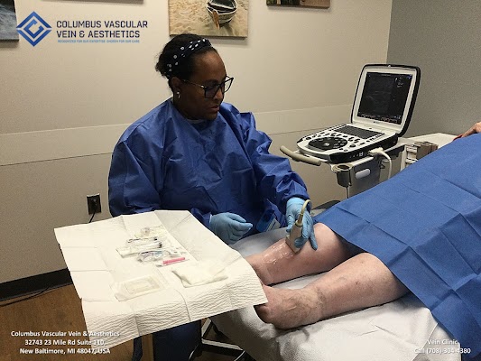
A standard sclerotherapy session begins with a focused assessment. For spider veins, we mark clusters with the patient standing. For visible varicose veins, or when symptoms suggest deeper issues, we review duplex ultrasound findings and decide on foam versus liquid and whether ultrasound guidance is needed. Compression stockings are measured and on hand.
The skin is cleaned thoroughly. For spider veins, we use very fine needles, often 30 or 32 gauge, and inject small amounts of sclerosant directly into the vessel. You may feel tiny pinches and a mild crampy sensation for a few seconds. For foam injections, we prepare the foam immediately before use to ensure consistency. Ultrasound guidance comes in when we target deeper reticular or varicose veins. After each injection, we apply gentle pressure and proceed along the planned map.
At the end, we place cotton or foam pads over treated areas, apply a compression stocking, and have you walk in the clinic for 10 to 20 minutes before departure. You go home with aftercare instructions and a follow-up plan. Most people schedule the next session at 4 to 8 weeks if more work is needed.
Short checklist: preparing and recovering well
- Bring your prescribed compression stockings and wear them as directed after your session. Avoid lotions or oils on your legs the day of the appointment. Plan for brisk walking after treatment, and avoid hot tubs, saunas, or high-intensity leg workouts for 48 hours. Protect treated areas from sun exposure to reduce pigmentation risk. Tell your clinician about prior clots, surgery, pregnancy, or medications like anticoagulants ahead of time.
What influences sclerotherapy effectiveness
Technique matters, but biology sets the stage. Patients with strong family histories of venous disease may grow new veins over time regardless of perfect treatment. Occupations that require long hours standing increase venous pressure, which can encourage recurrence. Weight, hormonal fluctuations, and connective tissue laxity also play a role.
For spider veins, treating feeders and using compression for one to two weeks can be the difference between partial and compelling results. For varicose veins, ensuring the deep drivers of reflux are addressed first preserves results.
Vein size dictates sclerosant concentration. Too weak, and the vein recanalizes. Too strong, and you risk skin injury, ulceration, or hyperpigmentation. There is no one-size-fits-all formula. An experienced practitioner adjusts concentration by anatomic zone. For instance, the ankle and shin often get gentler mixes than the outer thigh because the skin is thinner and arterial branches lie closer.
Risks, side effects, and how we minimize them
Most side effects are temporary and manageable. You can expect redness, mild swelling, and bruising. Matting, those fine pink webs near treated areas, occurs in a small percentage and often resolves with time or additional targeted injections. Hyperpigmentation appears as a brown track along the old vein path and fades in most patients within 3 to 12 months.
Trapped blood happens when a treated segment seals but retains deoxygenated blood. It looks like a tender, raised blue line. We often evacuate it with a tiny needle at follow-up, which speeds clearance and reduces pigmentation risk. Superficial phlebitis, a localized inflammatory reaction, presents as warmth and tenderness along a treated vein. Warm compresses, walking, compression, and anti-inflammatories usually resolve it quickly with guidance from your clinician.
Serious complications are rare with proper technique. Skin ulceration can occur if sclerosant leaks into tissue or if an arterial injection happens in a high-risk area. This is why ultrasound mapping and respect for anatomic no-go zones, especially around the ankle and foot, are essential. Allergic reactions are uncommon with modern agents like polidocanol, which is not histamine releasing, but any history of sensitivity should be disclosed.
Deep vein thrombosis is very uncommon in standard cosmetic sclerotherapy. Risk rises slightly with large-volume foam work, prior clotting history, immobility, or thrombophilia. We mitigate it by using the lowest effective volumes, encouraging early ambulation, and in select cases coordinating with the patient’s medical team.
Cost, sessions, and what “maintenance” really means
Sclerotherapy pricing varies by region and clinic. Many practices charge per session, often in the range of a few hundred dollars for spider veins, with higher pricing for ultrasound guided foam sessions for varicose veins due to the expertise and equipment involved. Insurance rarely covers cosmetic spider vein treatment. Coverage is more likely when there is documented venous insufficiency with symptoms, failed conservative measures, and a medical treatment plan for varicose veins.
Most patients with spider veins need 1 to 3 sessions per leg website area to reach their goals. If there are extensive networks, plan for staged treatments over several months. Varicose vein plans are more individualized. After treating the refluxing trunk with ablation or foam, some patients need one to two adjunct foam sessions or limited microphlebectomy for best contour.
Maintenance is not failure. Veins respond to life’s forces. If your work keeps you on your feet, or if hormones fluctuate, new spider veins may appear over years. A touch-up visit every year or two keeps the legs looking clear. Small reinvestments prevent the need for a major overhaul later.
Sclerotherapy vs laser, surgery, and other options
For leg spider veins, sclerotherapy usually outperforms transcutaneous laser. Laser may work for fine red telangiectasias and for patients who dislike needles, but it typically requires more sessions and can be less effective for blue reticulars. Combined approaches are common, using sclerotherapy for the network and a brief laser pass for resistant tiny red tips.

For varicose veins, compare sclerotherapy to surgery and thermal ablation. The old stripping operations have largely been replaced by endovenous laser and radiofrequency. These close the refluxing trunk with local anesthesia through a pinhole entry. Recovery is rapid, and the durability is excellent when the anatomy is favorable. Foam sclerotherapy is less invasive still, and it treats tortuous segments that wires and catheters cannot traverse. The trade-off is a somewhat higher chance of needing touch-ups.
Some ask about non-procedural options. Compression stockings, calf-strengthening, weight management, and elevation reduce symptoms and slow progression. They do not make existing varicose or spider veins disappear. Supplements and topical lotions do not collapse veins the way sclerotherapy does. That is not to dismiss lifestyle. Better circulation and leg health improve comfort and reduce recurrence risk after any vein treatment.

What to expect aftercare and healing time
After sclerotherapy, we recommend walking for 20 to 30 minutes right away and daily thereafter. Most people wear compression stockings during the day for 7 to 14 days after spider vein sessions and 2 to 3 weeks after foam for varicose veins, though exact protocols vary. You can shower, but avoid very hot baths, saunas, and high-intensity leg workouts for 48 hours to keep inflammation in check. Avoid heavy sun exposure on treated areas for a few weeks. If you tan easily, use clothing or high-SPF sunscreen to reduce pigmentation risk.
It is normal to feel mild itchiness at injection sites and firmness along treated varicose segments. These sensations fade over one to four weeks. If a tender lump appears, or if redness tracks more than expected, call your clinic. Prompt management speeds comfort and reduces visible marks.
Real-world scenarios that illustrate the nuances
A middle-aged nurse with scattered spider veins on the thighs and outer calves comes in between night shifts. We identify a few reticular feeders with transillumination and treat those first with a gentle liquid. Then we clear the overlying spider webs with micro sclerotherapy. She wears 20 to 30 mm Hg stockings during her shifts for two weeks, then as needed for swelling. At the six-week visit, most bruising has resolved, and we finish a few remaining strands in a short touch-up. Photos show a clear change, and she schedules a maintenance visit for the following year.
A contractor with bulging calf veins and evening heaviness presents after years on ladders. Ultrasound shows reflux in the Columbus Vascular Vein & Aesthetics in New Baltimore great saphenous vein and large tributaries. We plan endovenous ablation of the trunk, followed by ultrasound guided foam sclerotherapy for the tributaries two weeks later. He walks immediately after each session, wears compression for three weeks, and returns for a four-week scan to confirm closure. His symptoms improve first. The cosmetic softening follows over two to three months.
An avid runner with ankle telangiectasias and thin skin wants a cleaner look. We warn about slower clearance and higher matting risk near the ankle. We use a conservative concentration, tiny volumes, and meticulous pressure. Compression and sun avoidance are non-negotiable. Her results come in more slowly than thigh veins, but by three months she is pleased, and we avoid the matting that sometimes follows aggressive dosing.
The durability question: permanent or temporary results
Sclerotherapy permanently closes the specific treated veins. Those veins do not “reopen” in most cases if they were properly closed and not too large. What changes with time is the network around them. Genetics, hormones, and daily habits can bring new spider veins to the surface. The person sees “return of veins,” but these are different veins, not the same ones undoing the work. For varicose veins, if the underlying source of reflux is untreated, recanalization or outflow through collateral channels can create new bulges. That is why thorough evaluation and a stepwise plan matter.
Patients who view sclerotherapy as part of vein health, not just a one-off fix, get the best long-term satisfaction. They use compression for heavy days, keep moving, manage weight, and come in for quick touch-ups before small problems become large ones.
A short comparison at a glance
- Spider veins respond very well to liquid micro sclerotherapy, often 70 to 90 percent clearance with 1 to 3 sessions, best when feeders are addressed and compression is used. Reticular veins often feed spider clusters and should be treated first for stronger results. Varicose veins benefit from ultrasound guided foam sclerotherapy when used to treat tributaries or in conjunction with closing a refluxing trunk by thermal ablation or adhesive closure. Durability depends on treating the cause of reflux, tailoring sclerosant concentration, and following aftercare. Maintenance touch-ups every 1 to 3 years are common, especially for those with strong family history or standing occupations.
Final thoughts for selecting the right approach
A good sclerotherapy plan begins with the right diagnosis. If your primary concern is spider veins, ask your clinician whether there are reticular feeders that should be treated first, and what compression to wear after your session. If you have bulging varicose veins or symptoms like heaviness and swelling, request a duplex ultrasound. Discuss whether your saphenous veins show reflux and how best to address it, including the role of foam sclerotherapy versus laser or radiofrequency.
The sclerotherapy method is a versatile, minimally invasive treatment. Used thoughtfully, it offers fast, effective results with little downtime. The art lies in matching the technique to the vein type, respecting anatomy, and planning for the long game. That is how legs look better and feel better, not just for a season, but for years.